
3D Visualization in Biomedical Applications

The advances in medical imaging capabilities since 1970 have been developed, applied, and accepted at a volume and pace unprecedented in medical history. Computer and digital radiographic technology and techniques have significantly expanded the possibilities for accurate, quantitative, and noninvasive visualization and measurement of intracorporeal morphology and function. These advances have provided a variety of new diagnostic methodologies for clinical evaluation of health and disease. 3-D–imaging and –visualization methods are emerging as the methods of choice in many clinical examinations, replacing some previously routine procedures and significantly complementing others. The continuing evolution of 3-D and visualization imaging promises even greater capabilities for accurate noninvasive clinical diagnoses and treatment, as well as for quantitative biological investigations and scientific exploration, targeted at ever increasing our understanding of the human condition and how to improve it.
Individual molecules and cells, as well as various tissue and interstitial interfaces, as well as full organs, organ systems, and body sections, are all visualizable objects in biology and medicine. These items comprise biophysical, biomedical and physiological features, as well as functional attributes of these systems. With the introduction of high-resolution tomographic scanners and imaging systems, it is now possible to visualize such things and their functions in three dimensions. The direct, completely immersive, real-time multisensory merging of real and virtual information has the potential to revolutionize medical practice and biologic research. Accurate anatomy and function mapping, improved diagnosis, accurate treatment planning and rehearsal, and education/training are some of the medical applications. High-performance computers, improved image processing, and high-fidelity rendering capabilities have aided in making significant progress toward these objectives. With these advancements, three-dimensional visualization has various essential applications that will have a substantial impact on medical practice and biological research. Here are some applications.
Virtual Endoscopy
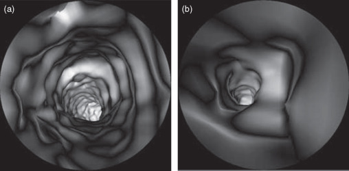
Virtual endoscopy is one of the most important clinical uses of 3-D visualization and volume rendering. Virtual endoscopy has the advantage of noninvasive cavity exploration, which could be beneficial in screening treatments. Three-dimensional computed endoscopic visualizations are created using two methods. The first method is direct volume rendering, while the second is to create volume models for real-time exploration of the region of interest.
Neurosurgery
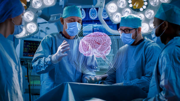
Neurosurgery is a difficult technique that necessitates a thorough understanding of the numerous interactions that exist between normal anatomy and disease. Preoperative multimodality imaging scanning is performed on patients with brain tumors, arterial-venous malformations, or other difficult internal brain pathologies to aid the neurosurgeon in understanding the anatomy of interest.
Prostate Cancer
It is common practice to surgically remove many cancerous prostates, even though subsequent pathological examination of the excised tissues suggests that some surgeries could have been avoided.
Cardiac and Coronary Artery Disease
There are significant evolving applications of 3-D interactive visualization in the treatment of heart and coronary artery disease shows unique visualizations of rendered reconstructions from the Mayo Dynamic Spatial Reconstructor.
Radiation Treatment Planning
Using CT and MRI fused scans, 3-D dosimetry can be computed for simulated treatment beams positioned to maximize dose to the target and minimize dose to critical structures in the treatment field.
Microscopy Imaging
The application of 3-D visualization and analysis techniques to the field of microscopy has grown significantly in recent years. These techniques have been successfully applied in light and electron microscopy, but the advent of confocal microscopy and other 3-D microscope modalities has led to the rapid growth of 3-D visualization of microscopic structures.
Author: Peula Thilakcha
Biomedical and Earth Science Division